pet-x-rays
Pet X-rays are medical diagnostic tools used to examine the internal organs and tissues of pets. Similar to human X-rays, they help veterinarians identify abnormal conditions such as bone injuries, tumors, cysts, and other issues.
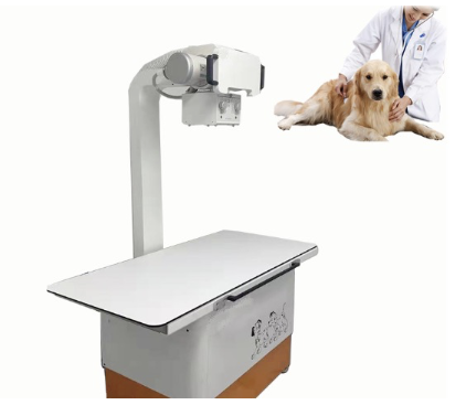
During a pet X-ray examination, the pet lies on a specially designed X-ray table and needs to remain still to avoid affecting the result. Prior to the examination, veterinarians will inquire about the pet's health status and medical history and explain the entire examination process and precautions to the owner.
There are generally two types of pet X-ray examinations: conventional film X-rays and digital X-rays. Conventional X-rays use traditional X-ray films to record images, while digital X-rays store images in electronic devices and can display and process images more quickly.
Although pet X-ray examinations are helpful for diagnosing pet health issues, there are also some potential risks. Frequent, long-term X-ray examinations may increase the risk of cancer in pets. Therefore, it is necessary to weigh the pros and cons and strictly control the frequency, minimizing the dosage when conducting X-ray examinations.
In conclusion, pet X-rays are important tools for veterinarians to diagnose internal illnesses in pets, but precautions need to be taken to minimize potential negative effects. If you have any questions regarding pet X-rays, please consult professional pet healthcare providers.
