Veterinary X-ray Machines Comprehensive Guide Technical Specifications and Clinical Applications
Veterinary X-ray Machines: A Comprehensive Guide for Modern Veterinary Practices
"The integration of advanced imaging technology has revolutionized veterinary diagnostics, allowing for non-invasive examination of internal structures with unprecedented clarity." - American Veterinary Medical Association
In the evolving landscape of veterinary medicine, diagnostic imaging stands as a cornerstone of modern practice. Veterinary X-ray machines have transitioned from basic radiographic tools to sophisticated digital systems that provide detailed insights into animal anatomy and pathology. This comprehensive guide explores the technical specifications, clinical applications, and practical considerations for implementing radiographic technology in veterinary settings.
Technical Evolution and Current Standards
The development of veterinary radiography has paralleled human medical advancements while addressing unique challenges in animal imaging. According to research published in the Journal of Veterinary Radiology & Ultrasound, digital radiography (DR) systems now dominate modern veterinary practices, offering significant advantages over traditional film-based systems. These include reduced radiation exposure, immediate image availability, and enhanced image manipulation capabilities.
Modern veterinary X-ray systems typically operate within the 40-150 kVp range, with mA settings adjustable based on patient size and anatomical region. The FDA's guidelines for veterinary X-ray systems emphasize the importance of proper calibration and regular quality assurance testing to ensure diagnostic accuracy and radiation safety.
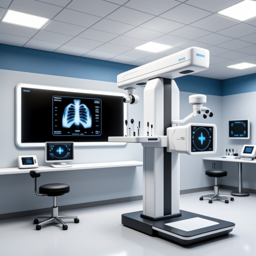
Key Technical Specifications
- Generator Power: 20-100 kW high-frequency generators for consistent output
- Focal Spot Size: Dual focal spots (0.6/1.2 mm) for detail and general radiography
- Detector Technology: Amorphous silicon or selenium flat panel detectors with pixel pitches of 100-200 μm
- Image Processing: Advanced algorithms for noise reduction and edge enhancement
- Compatibility: DICOM 3.0 standard for integration with practice management systems
Clinical Applications Across Species
Veterinary radiography serves diverse diagnostic purposes across multiple species. The Veterinary Information Network's clinical guidelines detail specific protocols for different animal categories:
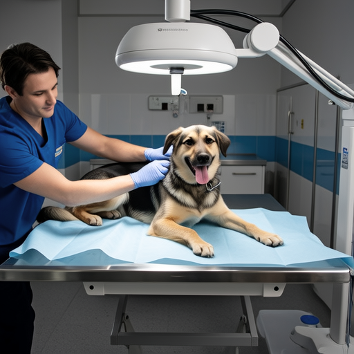
Small Animal Practice
Canine and feline radiography represents the most common application, with specialized techniques for orthopedic evaluation, thoracic imaging, and abdominal studies. Digital systems allow for rapid assessment of trauma cases, with studies showing diagnostic accuracy improvements of 15-20% compared to film systems for subtle fractures.
Equine and Large Animal Imaging
Portable and mobile X-ray units have transformed large animal practice, enabling field diagnostics for limb abnormalities, dental issues, and respiratory conditions. High-power generators (up to 100 kW) are essential for penetrating dense equine tissues, while specialized positioning devices accommodate anatomical variations.
Exotic and Zoo Animal Applications
Specialized techniques have been developed for avian, reptilian, and small mammal radiography, often requiring modified exposure settings and unique positioning approaches. The American Association of Equine Practitioners provides specific guidelines for non-traditional species imaging.
Digital Revolution in Veterinary Imaging
The transition from analog to digital radiography represents the most significant advancement in veterinary imaging history. According to market analysis by Grand View Research, the global veterinary imaging market is projected to reach $2.3 billion by 2027, driven primarily by digital technology adoption.
Direct vs. Indirect Digital Systems
Direct radiography (DR) systems utilize flat panel detectors that convert X-rays directly to electrical signals, offering superior image quality and workflow efficiency. Indirect systems employ scintillator layers that convert X-rays to light, which is then detected. While both systems offer advantages over computed radiography (CR), DR systems typically provide better detective quantum efficiency (DQE).
PACS Integration and Telemedicine
Modern veterinary practices increasingly implement Picture Archiving and Communication Systems (PACS) that integrate with practice management software. This enables efficient image storage, retrieval, and sharing for specialist consultation. The Veterinary Practice News reports that telemedicine consultations utilizing digital radiographs have increased by 300% since 2020.
Workflow Efficiency Comparison
Digital systems reduce examination time by 60-70% compared to film-based radiography, with immediate image availability eliminating chemical processing delays.
Diagnostic Accuracy Metrics
Studies indicate 12-18% improvement in diagnostic confidence with digital systems, particularly for subtle pulmonary patterns and early degenerative joint disease.
Cost-Benefit Analysis
While initial investment is higher, digital systems demonstrate ROI within 18-24 months through reduced consumable costs and increased patient throughput.
Radiation Safety and Regulatory Compliance
Veterinary radiographic practice requires strict adherence to radiation safety protocols. The International Atomic Energy Agency's radiation protection guidelines provide comprehensive safety standards for veterinary facilities.
Personnel Protection Measures
Lead aprons (minimum 0.5 mm Pb equivalent), thyroid shields, and protective gloves remain essential for staff safety. Modern facilities increasingly implement lead-lined control rooms with viewing windows, reducing occupational exposure by 95% compared to handheld shielding.
Patient Safety Considerations
Collimation to the area of interest, appropriate exposure settings, and use of positioning aids minimize patient radiation exposure. Studies published in Veterinary Radiology & Ultrasound demonstrate that digital systems can reduce patient dose by 30-50% while maintaining diagnostic quality.

Equipment Selection and Practice Integration
Selecting appropriate radiographic equipment requires careful consideration of practice needs, patient demographics, and financial constraints. The Veterinary Team Brief's equipment selection guide recommends evaluating systems based on five key criteria:
- Clinical Requirements: Match system capabilities to common procedures and patient types
- Technical Specifications: Evaluate generator power, detector size, and image processing features
- Workflow Integration: Assess compatibility with existing practice management systems
- Service and Support: Consider manufacturer reputation and local service availability
- Total Cost of Ownership: Calculate long-term expenses including maintenance and upgrades
Future Trends and Innovations
Emerging technologies continue to shape veterinary radiography. Artificial intelligence algorithms for automated image interpretation, wireless detector technology for enhanced mobility, and dose monitoring systems represent the next frontier. Research from Nature's veterinary science publications indicates that AI-assisted diagnosis could reduce interpretation time by 40% while improving detection of subtle abnormalities.
Key Takeaways for Veterinary Practices
Modern veterinary X-ray technology offers unprecedented diagnostic capabilities when properly implemented. Successful integration requires balancing technical specifications with practical workflow considerations, while maintaining rigorous safety standards. As digital technology continues to evolve, veterinary practices that strategically invest in radiographic equipment will enhance diagnostic accuracy, improve patient outcomes, and strengthen their competitive position in an increasingly technology-driven field.
For comprehensive equipment evaluation and implementation guidance, consult with veterinary radiologists and equipment specialists who can provide practice-specific recommendations based on current clinical standards and emerging technological developments.
