Veterinary X-ray Imaging Comprehensive Guide Technology Applications
The Art and Science of Veterinary X-ray Imaging: A Comprehensive Guide
Veterinary radiography has evolved from a niche diagnostic tool to an indispensable component of modern animal healthcare. This article explores the technical advancements, clinical applications, and future directions of veterinary X-ray imaging, drawing insights from leading veterinary institutions and research publications.
Historical Perspective and Technological Evolution
The journey of veterinary radiography began shortly after Wilhelm Röntgen's discovery of X-rays in 1895. According to the American Veterinary Medical Association, the first documented veterinary X-ray was performed in 1896 on a dog with a gunshot wound. Today, digital radiography systems have largely replaced traditional film-based methods, offering superior image quality and reduced radiation exposure.
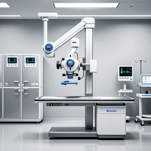
Figure 1: State-of-the-art digital radiography system in veterinary practice
Clinical Applications Across Species
Canine and Feline Radiography
Small animal radiography represents approximately 65% of all veterinary imaging procedures. Common indications include:
- Orthopedic evaluations for dysplasia and fractures
- Thoracic imaging for cardiac and respiratory conditions
- Abdominal studies for foreign body detection and organ assessment
- Dental radiography for periodontal disease evaluation
The American College of Veterinary Radiology emphasizes that proper positioning techniques are crucial for diagnostic accuracy, with sedation often required to minimize motion artifacts.
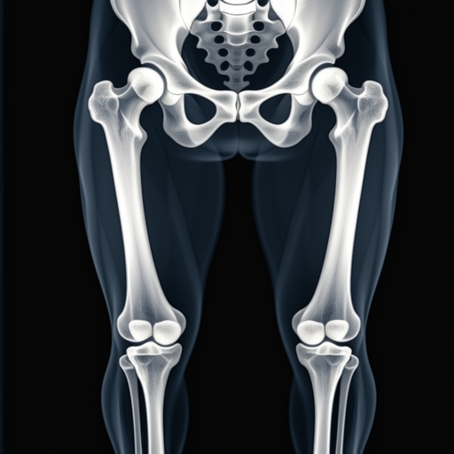
Figure 2: Radiographic assessment of canine hip joints
Equine and Large Animal Imaging
Large animal radiography presents unique challenges due to animal size and the need for portable equipment. According to research published in the International Veterinary Information Service, digital radiography has revolutionized equine practice by enabling:
- Immediate image availability in field settings
- Enhanced visualization of subtle bone changes
- Reduced retake rates through exposure feedback
Technical Considerations and Safety Protocols
Radiation Safety
Veterinary staff must adhere to ALARA principles (As Low As Reasonably Achievable). The Occupational Safety and Health Administration mandates regular monitoring with dosimeters and proper shielding implementation.
Image Quality Factors
Optimal radiographic quality depends on proper kVp/mA selection, collimation, and patient positioning. Digital systems offer post-processing advantages including window leveling and measurement tools.
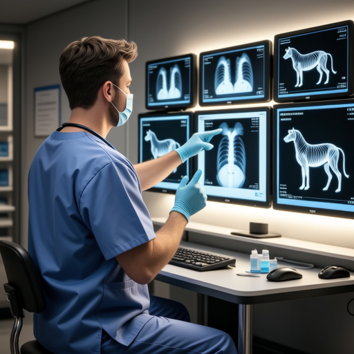
Figure 3: Digital image analysis in veterinary diagnostics
Emerging Technologies and Future Directions
Recent advancements documented by the Radiological Society of North America include:
- Cone-beam CT: Providing 3D imaging for dental and orthopedic applications
- Artificial Intelligence: Automated fracture detection and abnormality screening
- Tele-radiology: Remote consultation services expanding access to specialist interpretation
- Contrast-enhanced techniques: Improved soft tissue visualization
Economic Considerations
While digital systems require significant initial investment, studies show they reduce long-term costs through decreased film/chemical expenses and improved workflow efficiency. The Veterinary Practice News reports that practices typically recover digital equipment costs within 2-3 years through increased procedure volume and reduced retakes.
Educational and Training Requirements
Veterinary radiologists complete 4-5 years of specialized residency training following veterinary school. According to the AVMA Educational Commission, continuing education in radiation physics, anatomy, and pathology is essential for maintaining diagnostic proficiency. Many institutions now offer hybrid learning programs combining online coursework with hands-on workshops.
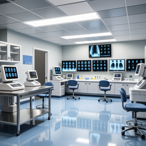
Figure 4: Modern veterinary imaging education facility
Conclusion
Veterinary radiography continues to advance through technological innovation and improved understanding of species-specific anatomy. As imaging modalities become more sophisticated and accessible, they will play an increasingly vital role in early disease detection, treatment planning, and improving outcomes across all veterinary species. The integration of AI and telemedicine promises to further democratize access to high-quality radiographic interpretation, ultimately benefiting animal welfare worldwide.
References: American Veterinary Medical Association, American College of Veterinary Radiology, International Veterinary Information Service, Radiological Society of North America, Veterinary Practice News. All links open in new windows for reader convenience.
