Veterinary CT Imaging Transforming Animal Healthcare Through Advanced Diagnostics
Veterinary CT: Revolutionizing Animal Diagnostics with Precision Imaging
In the evolving landscape of veterinary medicine, Computed Tomography (CT) has emerged as a transformative diagnostic tool, offering unparalleled insights into animal anatomy and pathology. Unlike traditional radiography, CT provides cross-sectional images that eliminate superimposition, allowing veterinarians to visualize structures in three dimensions with exceptional clarity.
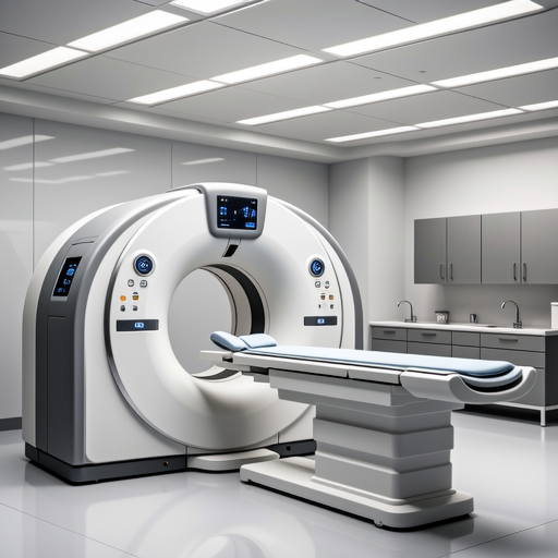
Modern veterinary CT scanner with advanced imaging capabilities
The Technical Foundations of Veterinary CT
Veterinary CT operates on the same fundamental principles as human CT scanners but incorporates adaptations for diverse species and sizes. The technology utilizes X-ray beams that rotate around the patient, with detectors measuring attenuation coefficients to reconstruct detailed anatomical slices. According to the American Veterinary Medical Association (AVMA), modern veterinary CT systems can achieve slice thicknesses as fine as 0.5mm, enabling detection of minute abnormalities that would remain invisible on conventional radiographs.
Key technical advancements include:
- Multi-detector arrays allowing rapid whole-body scans in seconds
- Low-dose protocols developed specifically for pediatric and exotic patients
- Dual-energy capabilities for tissue characterization and contrast enhancement
- Dynamic perfusion imaging for functional assessment of organs
Clinical Applications Across Species
Canine and Feline Medicine
In companion animal practice, CT has become indispensable for neurological assessments, oncological staging, and orthopedic evaluations. The American College of Veterinary Radiology (ACVR) reports that CT identifies 40% more nasal cavity tumors than radiography alone, significantly impacting treatment planning and prognosis.
Equine Diagnostics
For equine patients, standing CT systems have revolutionized lameness evaluation. These specialized units allow imaging of distal limbs without general anesthesia, reducing risks and recovery time. Research published in the Journal of Veterinary Radiology & Ultrasound demonstrates CT's superiority in detecting subtle navicular bone changes months before radiographic changes appear.
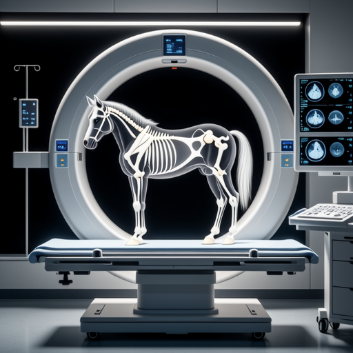
Standing CT examination of equine distal limb
Exotic and Wildlife Applications
The versatility of veterinary CT extends to exotic species, where anatomical knowledge is often limited. Zoological institutions utilize CT for:
- Pre-surgical planning for complex procedures in reptiles and birds
- Dental assessments in lagomorphs and rodents where malocclusion is common
- Conservation research studying anatomical adaptations in endangered species
- Forensic investigations in wildlife crime cases
The Wildlife Society notes that CT has become crucial for non-invasive study of internal structures in species where dissection is impractical or unethical.
Advancements in Imaging Protocols
Modern veterinary CT incorporates sophisticated protocols tailored to specific clinical questions:
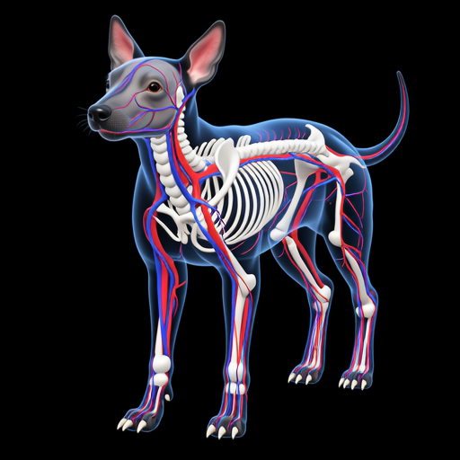
3D reconstruction of canine vascular anatomy using CT angiography
Safety Considerations and Radiation Protection
While CT delivers higher radiation doses than conventional radiography, modern systems incorporate multiple dose-reduction strategies:
ALARA Principle: All veterinary CT protocols follow the "As Low As Reasonably Achievable" principle for radiation exposure. Automatic exposure control adjusts tube current based on patient thickness, while iterative reconstruction algorithms maintain image quality at reduced doses.
The International Atomic Energy Agency (IAEA) provides guidelines for veterinary radiation safety, emphasizing that diagnostic benefits typically outweigh minimal risks when protocols are properly optimized.
Future Directions and Innovations
Emerging technologies promise to further enhance veterinary CT capabilities:
Artificial Intelligence Integration
Machine learning algorithms for automated lesion detection and measurement, reducing interpretation time and increasing consistency.
Portable CT Systems
Compact units for field use in large animal practice and disaster response situations.
Functional Imaging
Combined PET-CT systems entering veterinary markets for molecular imaging applications.
Conclusion
Veterinary CT has evolved from a specialized tool to a cornerstone of modern animal diagnostics. Its ability to provide detailed, three-dimensional anatomical information has transformed clinical decision-making across species. As technology advances and becomes more accessible, CT will continue to push the boundaries of what's possible in veterinary medicine, improving outcomes for patients from companion animals to wildlife conservation subjects.
References: American Veterinary Medical Association, American College of Veterinary Radiology, Journal of Veterinary Radiology & Ultrasound, Wildlife Society, International Atomic Energy Agency
