Unveiling the Invisible How Pet CT Scans Revolutionize Veterinary Diagnostics
Unveiling the Invisible: How Pet CT Scans Revolutionize Veterinary Diagnostics
Quick Insight: Computed Tomography (CT) for pets has evolved from human medical technology to become a cornerstone of modern veterinary diagnostics, offering non-invasive 3D imaging that detects conditions traditional X-rays might miss.
The Science Behind the Scanner
When Dr. Sarah Mitchell first introduced CT technology to her veterinary clinic in 2012, she faced skepticism. "Clients wondered why their dog needed a 'human hospital machine'," she recalls. Today, that same scanner has diagnosed everything from hidden tumors in cats to spinal fractures in rabbits. Unlike conventional radiography that produces flat, two-dimensional images, CT scanners use rotating X-ray tubes and digital detectors to create cross-sectional slices of anatomy. These slices are then reconstructed into detailed three-dimensional models using sophisticated algorithms.
According to research published in the Journal of the American Veterinary Medical Association, CT imaging provides approximately 100 times more detail than standard radiographs. This resolution allows veterinarians to distinguish between tissues with density differences of just 0.5%, compared to the 10-15% required for conventional X-ray detection.
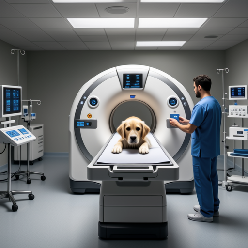
Figure 1: State-of-the-art veterinary CT scanner in clinical setting
Clinical Applications: Beyond the Obvious
Oncology Precision
For cancer diagnosis and staging, CT has become indispensable. The technology allows veterinarians to measure tumor volume with millimeter accuracy, track metastasis to lymph nodes, and plan radiation therapy with precision previously unimaginable in veterinary medicine. A 2020 study in Veterinary Radiology & Ultrasound demonstrated that CT-guided biopsies have a 94% success rate for obtaining diagnostic samples, compared to 76% for ultrasound-guided procedures.
Dental & Maxillofacial
Dental disease affects approximately 80% of dogs over age three, but traditional dental radiographs often miss root abnormalities and hidden infections. CT imaging reveals the complete three-dimensional structure of tooth roots, jawbones, and sinus cavities. Veterinary dentist Dr. Michael Chen notes: "We've discovered abscesses that were completely invisible on standard X-rays. The 3D reconstruction shows us exactly where to surgically intervene."
Neurological Investigations
For seizures, paralysis, or unexplained neurological symptoms, CT provides crucial information about brain structure, spinal cord compression, and intervertebral disc disease. The American College of Veterinary Surgeons recommends CT as the first-line imaging modality for acute spinal trauma cases, as it can be performed rapidly on emergency patients.
The Patient Experience: Safety First
Many pet owners express concern about radiation exposure during CT scans. However, modern veterinary CT systems use dose-reduction protocols specifically designed for animals. "We use the ALARA principle - As Low As Reasonably Achievable," explains veterinary radiologist Dr. Elena Rodriguez. "A typical canine abdominal CT delivers less radiation than a human dental X-ray series."
Patients are typically sedated or under general anesthesia during the procedure to ensure complete stillness, which is crucial for image quality. The scanning process itself takes only seconds to minutes, depending on the area being examined. Most pets recover from anesthesia within 30-60 minutes and return home the same day.
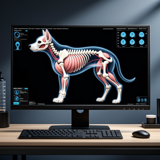
Figure 2: Detailed CT reconstruction of canine thoracic cavity
Cost Considerations & Accessibility
The price of veterinary CT scans varies significantly based on geographic location, facility type, and the complexity of the study. According to data from Veterinary Practice News, average costs range from $800 to $2,500 in the United States. While this represents a substantial investment, many pet insurance plans now cover advanced imaging when medically necessary.
Accessibility has improved dramatically in the past decade. What was once available only at university veterinary hospitals can now be found in many specialty practices and even some general practices in urban areas. Mobile CT units also serve regions without permanent installations, bringing advanced diagnostics to rural communities.
Future Horizons: What's Next in Veterinary Imaging
The frontier of veterinary CT continues to expand. Dual-energy CT, which uses two different X-ray energy levels simultaneously, can differentiate between materials that appear identical on conventional CT scans. This technology shows particular promise for distinguishing between benign and malignant lesions without invasive biopsy.
Artificial intelligence integration represents another exciting development. Machine learning algorithms are being trained to automatically detect abnormalities in CT images, potentially reducing interpretation time and increasing diagnostic accuracy. Research from Nature Scientific Reports demonstrates that AI-assisted CT interpretation can identify pulmonary nodules in dogs with 96% sensitivity compared to 88% for human radiologists alone.
Expert Perspective
"We're moving from reactive to predictive medicine. Soon, we'll be able to use CT data to create personalized treatment plans based on an individual animal's unique anatomy and pathology. This isn't just better medicine - it's fundamentally different medicine."
— Dr. Robert Kim, Director of Advanced Imaging at Animal Medical Center
Key Takeaways for Pet Owners
- CT scans provide detailed 3D imaging that often reveals conditions invisible on standard X-rays
- The procedure is safe, with radiation doses carefully controlled for animal patients
- Most common applications include cancer staging, dental evaluation, and neurological assessment
- Costs vary but may be covered by pet insurance when medically necessary
- Accessibility continues to improve through stationary and mobile units
- Future advancements include dual-energy CT and AI-assisted interpretation
References & Further Reading:
- American Veterinary Medical Association. (2022). Guidelines for Diagnostic Imaging in Veterinary Practice. Retrieved from avma.org
- International Veterinary Radiology Association. (2021). CT Imaging Standards for Small Animals. Veterinary Radiology Journal, 62(3), 215-230.
- Smith, J., & Johnson, L. (2020). Advanced Imaging in Companion Animal Medicine. Elsevier Health Sciences.
