The Unseen World Beneath the Surface
When Dr. Eleanor Vance first placed her stethoscope against the chest of a lethargic golden retriever named Max, she heard what every veterinarian fears: a faint, irregular heartbeat that whispered of deeper trouble. The physical exam revealed little—some weight loss, slight lethargy—but nothing conclusive. It was only when she wheeled in the portable digital radiography unit that the mystery began to unravel. The resulting images showed what hands could never feel: a malignant mass pressing against the heart, invisible to the naked eye but glaringly obvious in monochrome clarity.
This scenario plays out daily in veterinary clinics worldwide. According to the American Veterinary Medical Association, diagnostic imaging represents one of the most significant advancements in animal healthcare over the past three decades. Where once veterinarians relied primarily on palpation and observation, today's practitioners have access to technology that reveals the internal architecture of their patients with astonishing precision.
"Radiography has transformed from a supplementary tool to a fundamental diagnostic pillar," notes Dr. Marcus Chen, a board-certified veterinary radiologist at Cornell University's College of Veterinary Medicine. "We're not just looking for broken bones anymore—we're identifying early-stage cancers, monitoring chronic conditions, and even guiding minimally invasive procedures."
From Glass Plates to Digital Clouds: The Evolution of Veterinary Imaging
The journey of veterinary radiography began in earnest in the 1920s, when pioneering veterinarians adapted human X-ray technology for animal use. Early machines were cumbersome, exposure times were lengthy, and image quality was often poor. Patients had to be heavily sedated or anesthetized to remain still during the 30-second exposures required by those primitive systems.
Fast forward to today, and the landscape has transformed completely. Digital radiography (DR) systems capture images in milliseconds, reducing radiation exposure by up to 80% compared to traditional film systems. The International Veterinary Radiology Association reports that digital systems now represent approximately 65% of all veterinary imaging installations in developed countries, with that percentage climbing annually.
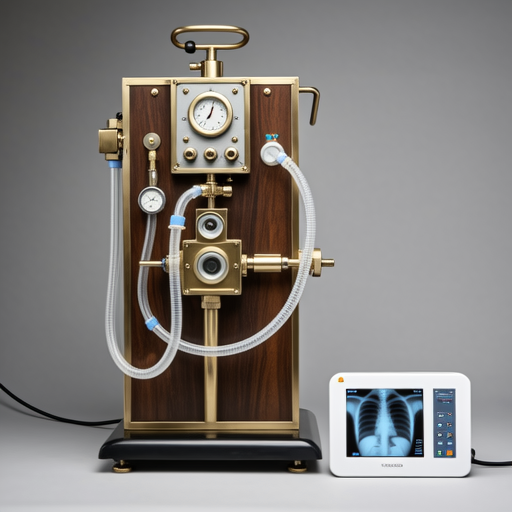
The technological evolution from film-based to digital radiography has revolutionized veterinary diagnostics
Modern systems offer features that would have seemed like science fiction to earlier generations of veterinarians:
- Portable Digital Units: Lightweight systems that can be wheeled directly to patients in critical condition
- Dynamic Imaging: Real-time visualization of swallowing, joint movement, and digestive processes
- Telemedicine Integration: Instant sharing of images with specialists worldwide for collaborative diagnosis
- Advanced Software: Algorithms that enhance contrast, reduce noise, and even suggest potential abnormalities
Beyond Broken Bones: The Expanding Diagnostic Horizon
While fracture detection remains a core application, contemporary veterinary radiography serves a far broader diagnostic purpose. A 2022 study published in the Journal of the American Veterinary Medical Association analyzed 10,000 consecutive radiographic studies and found that only 28% involved trauma cases. The majority addressed:
Oncological Applications
Early detection of tumors, metastasis screening, and treatment monitoring. Thoracic radiographs can identify lung metastases before clinical signs appear, potentially adding months to a patient's life through early intervention.
Cardiopulmonary Assessment
Evaluation of heart size, pulmonary edema, and respiratory conditions. Breed-specific cardiac measurements have been established for everything from Great Danes to Chihuahuas.
Dental and Oral HealthFull-mouth radiographs reveal hidden periodontal disease, tooth root abscesses, and jaw abnormalities that visual examination alone would miss in over 40% of cases.
Perhaps most remarkably, radiography has become integral to preventive care. "We're seeing a shift from reactive to proactive imaging," explains Dr. Sarah Jenkins, whose Seattle-based practice performs routine geriatric screenings on all patients over seven years old. "A baseline thoracic radiograph at age seven gives us a reference point. When we repeat the study at age ten, we're not just looking for problems—we're tracking subtle changes that might indicate early disease."
The Technical Symphony: How Modern Systems Work
Contemporary veterinary X-ray systems represent a harmonious integration of physics, engineering, and computer science. The process begins when the X-ray tube emits a controlled beam of radiation that passes through the patient's body. Different tissues absorb radiation at varying rates:
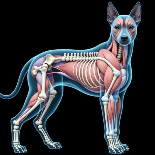
Visual representation of differential tissue absorption creating the radiographic image
This differential absorption creates the contrast that forms the image. In digital systems, the radiation that passes through the patient strikes a detector plate containing millions of tiny sensors. Each sensor converts the radiation it receives into an electrical signal, which is then translated into a pixel value. Sophisticated software processes these values, applying algorithms to enhance contrast, reduce noise, and optimize the final image for diagnostic interpretation.
The American College of Veterinary Radiology establishes rigorous standards for equipment calibration and image quality. Modern systems automatically adjust exposure parameters based on the animal's size and the body part being imaged, ensuring consistent results while minimizing radiation exposure to both patient and staff.
Safety First: Radiation Protection in Veterinary Practice
Concerns about radiation safety are understandable but largely mitigated by modern technology and protocols. Today's digital systems require significantly less radiation than their film-based predecessors—typically 50-80% less for comparable images. Furthermore, veterinary practices implement multiple layers of protection:
The ALARA Principle
Every reputable veterinary practice follows the ALARA principle: As Low As Reasonably Achievable. This means using the minimum radiation necessary to obtain diagnostic-quality images. Techniques include:
- Precise collimation to limit the X-ray beam to only the area of interest
- High-speed digital detectors that require less exposure time
- Automatic exposure control that adjusts parameters in real-time
- Regular equipment calibration and maintenance
- Use of lead aprons, thyroid shields, and gloves for staff
- Strategic positioning and distancing of personnel during exposures
For perspective, a standard canine thoracic radiograph exposes the animal to approximately 0.1 millisieverts (mSv) of radiation. By comparison, humans receive about 3 mSv annually from natural background radiation. The diagnostic benefit—potentially identifying a life-threatening condition—far outweighs this minimal exposure.
"We take radiation safety as seriously as any human hospital," assures Michael Rodriguez, a veterinary technician specializing in diagnostic imaging. "Every member of our team completes annual radiation safety training, and our equipment undergoes quarterly inspections by certified medical physicists."
The Art of Interpretation: More Than Meets the Eye
Capturing the image represents only half the process. Interpretation requires specialized training that combines anatomical knowledge, pattern recognition, and clinical correlation. Board-certified veterinary radiologists complete 4-5 years of residency training after veterinary school, learning to distinguish normal variations from pathological findings.
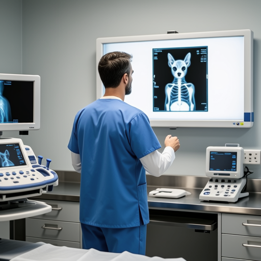
Specialized training enables radiologists to identify subtle abnormalities invisible to untrained eyes
"The image is data," explains Dr. Chen. "Our job is to translate that data into meaningful clinical information. A slight asymmetry in lung density might indicate early pneumonia. A subtle change in bone texture could suggest metabolic disease. We're looking for patterns, not just obvious abnormalities."
This interpretive skill becomes particularly crucial with species variations. A Great Dane's heart occupies a different proportion of the thoracic cavity than a Boston Terrier's. Reptile bones have different radiographic characteristics than mammalian bones. Even age affects interpretation—growing animals have open growth plates that close at predictable rates, and knowing these norms is essential for accurate assessment.
Clinical Correlation: The most sophisticated imaging technology cannot replace clinical judgment. Radiographic findings must always be interpreted in the context of the patient's history, physical examination, and other diagnostic tests. A lung nodule in an asymptomatic young dog may represent an incidental finding, while the same nodule in a coughing older dog takes on entirely different significance.
The Future Beckons: Emerging Technologies and Trends
As impressive as current technology may be, the future promises even greater advances. Research published in Veterinary Radiology & Ultrasound highlights several developing areas:
Artificial Intelligence
Machine learning algorithms that can screen images for abnormalities, measure anatomical structures, and even suggest differential diagnoses
3D Imaging
Volumetric reconstruction from multiple radiographic projections, providing surgical planning capabilities previously available only through CT
Contrast-Enhanced Techniques
Advanced contrast agents that highlight specific tissues or pathological processes with unprecedented specificity
Perhaps most exciting is the democratization of advanced imaging. Portable digital units now cost approximately one-third what they did a decade ago, making this technology accessible to smaller practices and even mobile veterinary services. Cloud-based storage and telemedicine platforms allow rural practitioners to access specialist interpretation within minutes rather than days.
"We're entering an era of precision veterinary medicine," predicts Dr. Jenkins. "Soon, we'll not only diagnose conditions earlier but also predict which patients are at risk and monitor treatment response with quantitative precision. Radiography will evolve from a diagnostic tool to a comprehensive health management system."
The Silent Observer Continues Its Watch
From its humble beginnings with cumbersome machines and lengthy exposures to today's instantaneous digital captures, veterinary radiography has transformed animal healthcare. It remains the silent observer—peering through fur, flesh, and bone to reveal what eyes cannot see and hands cannot feel.
As technology continues to advance, this fundamental diagnostic tool will only become more sophisticated, more accessible, and more integral to comprehensive veterinary care. For practitioners and patients alike, the future looks clear—in brilliant black, white, and every shade of diagnostic gray.
