The Evolution and Precision of X-ray Patient Positioning Systems
The Evolution and Precision of X-ray Patient Positioning Systems
In the realm of diagnostic imaging, the accuracy of an X-ray image begins long before the machine is activated. It starts with how the patient is positioned. Over the decades, X-ray patient positioners have evolved from simple manual aids to sophisticated, automated systems that are fundamental to modern radiology departments. This article explores their development, technological advancements, and critical role in ensuring diagnostic quality and patient safety.
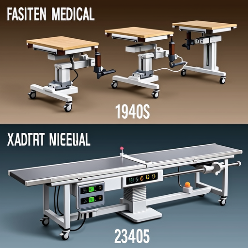
Historical progression of patient positioning technology in radiography.
From Manual Adjustments to Digital Precision
In the early days of radiography, technologists relied on wooden blocks, sandbags, and their own expertise to position patients. Consistency was a challenge, often leading to retakes and increased radiation exposure. The introduction of mechanical positioners in the mid-20th century marked a significant leap. These devices, often featuring adjustable arms, angle indicators, and locking mechanisms, brought a new level of reproducibility to radiographic exams.
Today, digital integration has transformed positioning into a precise science. Modern systems, such as those highlighted by the Radiological Society of North America (RSNA), incorporate motorized movements, touchscreen controls, and even AI-assisted alignment suggestions. These advancements minimize human error and optimize imaging protocols for specific anatomical regions.
Key Components and Functionality
A contemporary X-ray patient positioner typically consists of several integrated components:
- Motorized Table/Bucky Stand: Allows vertical and horizontal movement with precise digital controls, reducing physical strain on technologists.
- Angulation Mechanisms: Enable tilting for specialized views like Trendelenburg or Fowler's position, crucial for certain clinical scenarios.
- Immobilization Devices: Include straps, pads, and head holders that secure patients comfortably while preventing motion artifacts.
- Laser Guidance Systems: Project alignment markers onto the patient's body, ensuring accurate centering relative to the X-ray tube and detector.
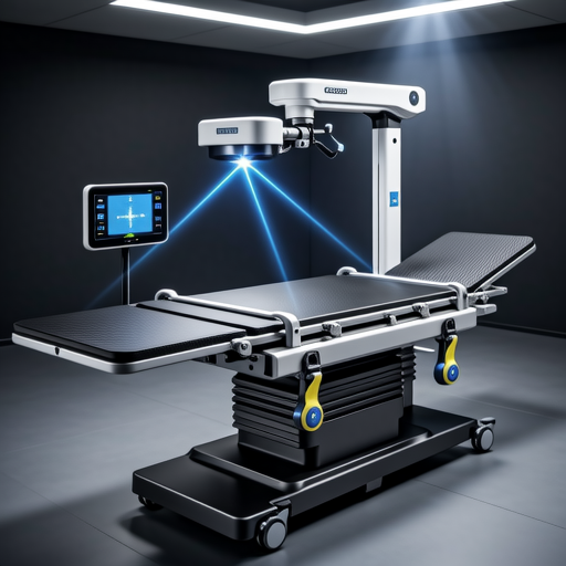
Modern motorized positioning system with integrated laser alignment.
Impact on Diagnostic Accuracy and Workflow
Proper positioning is directly linked to image quality. Misalignment can obscure pathologies, lead to false negatives, or necessitate repeat exposures. According to resources from the American Association of Physicists in Medicine (AAPM), consistent positioning reduces dose variability and enhances the reliability of longitudinal studies, where comparing images over time is essential.
In busy clinical environments, efficient positioning systems streamline workflow. Motorized adjustments cut setup times, while preset protocols allow quick switching between exam types. This efficiency not only boosts department throughput but also improves patient experience by reducing wait times and discomfort.
Safety and Ergonomics: A Dual Focus
Modern positioners prioritize safety for both patients and staff. Features like weight capacity indicators, obstacle detection sensors, and emergency stop buttons prevent accidents. For patients with mobility issues or trauma, specialized positioners facilitate imaging without exacerbating injuries.
Ergonomically, these systems protect technologists from musculoskeletal injuries. The Occupational Safety and Health Administration (OSHA) notes that reducing manual handling in healthcare settings decreases injury rates. Motorized tables eliminate the need for heavy lifting, while adjustable heights allow technologists to work in neutral postures.
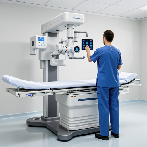
Ergonomic design reduces physical strain on radiology technologists.
Future Trends and Innovations
The future of patient positioning is leaning towards greater automation and intelligence. Emerging technologies include:
- AI-Driven Positioning: Systems that use machine learning to analyze patient anatomy and suggest optimal positioning, potentially integrating with electronic health records for personalized protocols.
- Robotic Assistants: Robotic arms that can gently reposition limbs or torsos with sub-millimeter accuracy, particularly useful in interventional radiology.
- Augmented Reality (AR) Guides: AR glasses that overlay positioning instructions directly into the technologist's field of view, hands-free.
- Enhanced Connectivity: IoT-enabled positioners that communicate with other imaging devices to automate entire exam sequences.
These innovations, discussed in forums like the RSNA's annual meeting, promise to further reduce human error, customize patient care, and push the boundaries of imaging precision.
Conclusion: Positioning as a Cornerstone of Radiology
X-ray patient positioners have transitioned from rudimentary tools to integral components of diagnostic imaging. Their evolution mirrors broader trends in healthcare technology: a shift towards precision, safety, and efficiency. As imaging demands grow and technology advances, these systems will continue to play a pivotal role in delivering accurate diagnoses and enhancing patient outcomes. Investing in advanced positioning technology is not just about upgrading equipment—it's about committing to higher standards of care.
References & Further Reading: This article draws on technical guidelines and research from authoritative sources including the Radiological Society of North America (RSNA), American Association of Physicists in Medicine (AAPM), and Occupational Safety and Health Administration (OSHA). For the latest advancements, peer-reviewed journals and industry conferences remain invaluable resources.
