The Evolution of Veterinary Imaging
Veterinary medicine has undergone a remarkable transformation over the past two decades, particularly in diagnostic imaging. What began with basic radiographic techniques has evolved into sophisticated digital systems requiring precise patient positioning. According to the American Veterinary Medical Association, diagnostic imaging now accounts for approximately 30% of all veterinary diagnostic procedures, with radiography maintaining its position as the most frequently utilized modality.
The challenge has always been obtaining consistent, high-quality images while ensuring patient safety and minimizing stress. Traditional methods involving manual restraint and makeshift positioning devices often resulted in compromised images, repeated exposures, and increased radiation exposure for both patients and staff.
What Exactly is a Veterinary X-ray Patient Positioner?
A veterinary X-ray patient positioner is a specialized device designed to accurately position animals for radiographic examinations. Unlike human medicine where patients can follow instructions, veterinary radiography requires equipment that can accommodate various species, sizes, and temperaments while maintaining precise anatomical alignment.
Modern positioners typically feature:
- Adjustable platforms with radiolucent surfaces
- Modular positioning aids (foam wedges, sandbags, troughs)
- Secure restraint systems that minimize stress
- Measurement guides for accurate centering
- Compatibility with both digital and traditional X-ray systems
The American College of Veterinary Radiology emphasizes that proper positioning is not merely about convenience—it's fundamental to diagnostic accuracy. Even minor positioning errors can lead to misinterpretation of findings, potentially affecting treatment decisions.
Technical Specifications and Design Considerations
When evaluating veterinary positioners, several technical factors determine their effectiveness:
Radiolucency: The platform material must not interfere with X-ray penetration. High-density polyethylene and carbon fiber composites have become industry standards, offering excellent radiolucency while maintaining structural integrity.
Load Capacity: Veterinary practices need equipment that can accommodate everything from a 2kg cat to a 70kg dog. Premium positioners typically support 150kg+ with stability.
Adjustability: Height adjustment mechanisms allow for proper alignment with X-ray tube heads, while tilt capabilities facilitate specialized views without repositioning the patient.
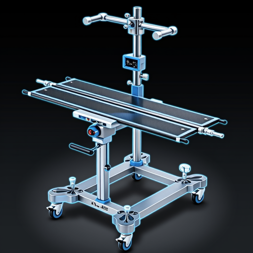
Modern veterinary positioner with adjustable components and measurement guides
A study published in the Journal of Veterinary Radiology & Ultrasound demonstrated that using dedicated positioning equipment reduced retake rates by 68% compared to manual positioning methods. This translates to significant time savings, reduced radiation exposure, and improved diagnostic confidence.
Clinical Applications Across Veterinary Specialties
The versatility of modern positioners makes them invaluable across various veterinary disciplines:
Orthopedic Imaging
Precise limb positioning is critical for evaluating fractures, joint diseases, and developmental conditions. Positioners with specialized limb holders and angle guides enable consistent orthogonal views essential for surgical planning.
Thoracic Radiology
Consistent positioning eliminates rotation artifacts that can mimic or mask pulmonary pathology. Dedicated thoracic positioners ensure proper sternal recumbency and symmetrical positioning for accurate cardiac and pulmonary assessment.
Exotic Animal Medicine
Specialized positioners with adjustable compartments and miniature positioning aids accommodate rabbits, reptiles, birds, and other exotic species that require unique positioning approaches.
The Veterinary Practice News reports that clinics implementing comprehensive positioning systems see a 40% reduction in anesthesia time for radiographic procedures, as precise positioning reduces the need for multiple attempts and adjustments.
Economic and Workflow Considerations
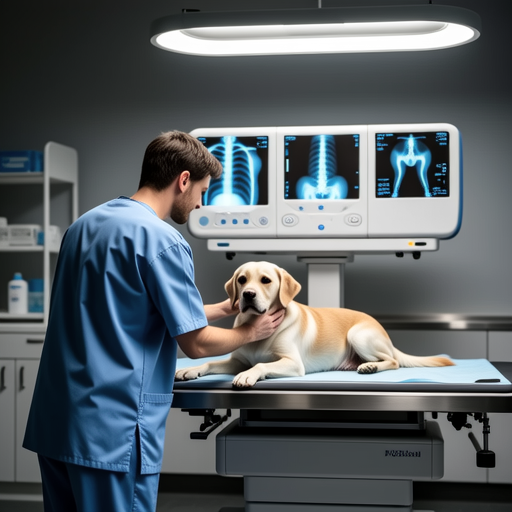
Efficient workflow with dedicated positioning equipment in veterinary practice
While the initial investment in quality positioning equipment may seem substantial, the return on investment becomes apparent through multiple channels:
Reduced Retakes: Each retake represents wasted time, materials, and radiation exposure. Positioners dramatically decrease retake rates.
Staff Efficiency: Technicians can position patients more quickly and consistently, increasing throughput.
Diagnostic Confidence: Higher quality images lead to more accurate diagnoses, reducing the need for additional imaging studies.
Staff Safety: Reduced manual handling decreases injury risk, while proper positioning minimizes radiation scatter exposure.
According to data from the Washington State University Veterinary Clinical Sciences Radiology Department, clinics using dedicated positioning systems report an average time savings of 8-12 minutes per radiographic study. For a busy practice performing 15-20 radiographs daily, this translates to 2-4 additional hours of productive time each week.
Future Developments in Veterinary Positioning Technology
The future of veterinary patient positioning is moving toward greater integration and automation:
Digital Integration
Positioners with built-in digital measurement systems that communicate directly with X-ray generators to automatically calculate exposure settings based on patient size and position.
AI-Assisted Positioning
Systems using artificial intelligence to analyze patient anatomy and suggest optimal positioning parameters for specific diagnostic questions.
Species-Specific Designs
Modular systems with interchangeable components optimized for different animal families, from canine/feline to equine and exotic species.
Industry analysts at Veterinary Practice News predict that within five years, integrated positioning systems will become standard in progressive veterinary practices, much as digital radiography replaced film-based systems over the past decade.
Implementing Positioning Systems in Your Practice
Transitioning to dedicated positioning equipment requires careful planning:
- Assess Your Needs: Consider the species you treat, volume of radiographs, and types of studies most frequently performed.
- Evaluate Space: Ensure your radiographic suite can accommodate the equipment while maintaining safe working distances.
- Staff Training: Proper utilization requires training on positioning principles and equipment operation.
- Quality Assurance: Establish protocols for regular equipment maintenance and positioning technique reviews.
- Gradual Implementation: Consider starting with basic positioning aids and expanding as your team becomes proficient.
The AVMA recommends that practices developing their imaging capabilities consult with veterinary radiologists or experienced technicians when selecting positioning equipment to ensure it meets their specific clinical needs.
The Bottom Line
Veterinary X-ray patient positioners represent more than just convenience equipment—they are fundamental tools for diagnostic excellence. By ensuring consistent, accurate positioning, these systems improve image quality, enhance diagnostic confidence, increase workflow efficiency, and promote radiation safety. As veterinary medicine continues to advance, investing in proper positioning technology remains one of the most impactful decisions a practice can make for both patient care and operational success.
