Comprehensive Guide to Veterinary X-ray Technology and Clinical Applications
The Art and Science of Veterinary X-ray Imaging
"In veterinary medicine, X-ray technology serves as our window into the unseen world of animal anatomy—revealing fractures, foreign bodies, and pathologies that would otherwise remain hidden." - Dr. Sarah Mitchell, American Veterinary Medical Association
Veterinary radiography has evolved from a niche diagnostic tool to an indispensable component of modern animal healthcare. What began with Wilhelm Röntgen's discovery of X-rays in 1895 has transformed into sophisticated digital systems capable of capturing detailed anatomical images in seconds. Today, veterinary clinics worldwide rely on radiographic imaging to diagnose conditions ranging from simple fractures to complex internal disorders.
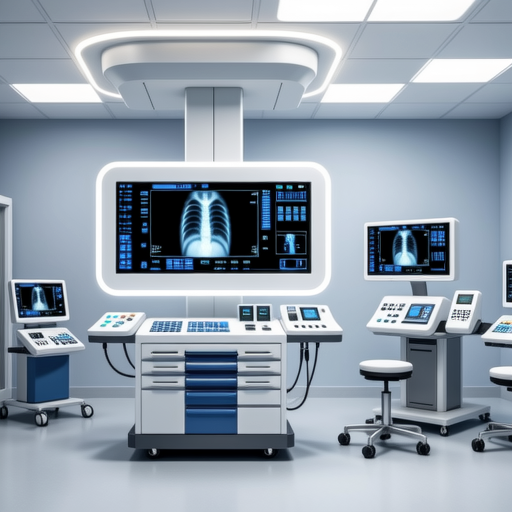
Figure 1: State-of-the-art digital radiography system in veterinary practice
Technical Evolution in Veterinary Radiology
The transition from film-based to digital radiography represents one of the most significant advancements in veterinary imaging. According to research published in the Journal of Veterinary Radiology & Ultrasound, digital systems offer several advantages including reduced radiation exposure, immediate image availability, and enhanced diagnostic capabilities through post-processing techniques.
Modern veterinary X-ray systems typically operate between 40-150 kVp (kilovolt peak) with exposure times ranging from 0.01 to 0.5 seconds, depending on the animal's size and the anatomical region being examined. The American College of Veterinary Surgeons emphasizes proper technique selection to optimize image quality while minimizing radiation dose.
Digital Radiography (DR)
Direct digital capture systems using flat-panel detectors provide immediate image acquisition with excellent spatial resolution. These systems typically offer 12-16 bit depth, allowing visualization of subtle tissue differences.
Computed Radiography (CR)
Phosphor plate systems that require separate processing but offer flexibility in positioning and cost-effectiveness for smaller practices. CR systems maintain diagnostic quality while being more affordable than DR alternatives.
Clinical Applications and Diagnostic Protocols
Veterinary radiography serves multiple diagnostic purposes across various specialties. Orthopedic evaluations represent approximately 45% of all veterinary radiographic studies, followed by thoracic imaging (30%) and abdominal studies (25%), according to data from the UC Davis Veterinary Medical Teaching Hospital.
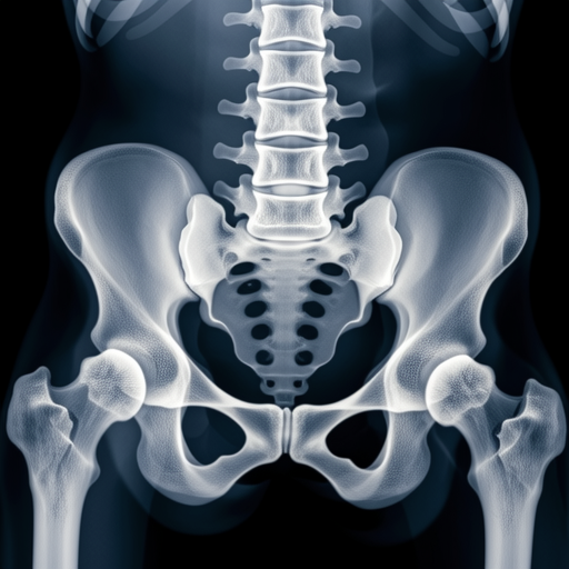
Figure 2: Radiographic assessment of canine hip joints for dysplasia evaluation
Standard Positioning Techniques
Proper patient positioning remains crucial for diagnostic accuracy. The Veterinary Information Network outlines standardized views including:
- Orthogonal Views: Minimum of two perpendicular projections (typically lateral and ventrodorsal/dorsoventral)
- Stress Views: Used for ligamentous injury assessment in joints
- Contrast Studies: Employing barium or iodine-based agents for gastrointestinal or urinary tract visualization
Safety Protocols and Radiation Protection
Radiation safety represents a paramount concern in veterinary practice. The International Atomic Energy Agency establishes guidelines for veterinary radiation protection, emphasizing the ALARA principle (As Low As Reasonably Achievable).
Essential Safety Measures
- Lead aprons (minimum 0.5mm lead equivalence) for all personnel in radiographic suite
- Thyroid shields and leaded glasses for frequent operators
- Dosimetry monitoring for staff exceeding 10% of occupational limits
- Proper collimation to restrict radiation field to area of interest
- Regular equipment calibration and quality assurance testing
Interpretation Challenges and Diagnostic Pitfalls
Veterinary radiographic interpretation requires specialized knowledge of comparative anatomy across species. A study in Veterinary Radiology & Ultrasound identified common interpretation errors including:
Technical Artifacts
Motion blur, improper exposure, grid lines, and processing errors can mimic or obscure pathology.
Anatomical Variants
Normal breed variations and age-related changes misinterpreted as pathological findings.
Projection Errors
Rotation or obliquity creating false impressions of asymmetry or displacement.
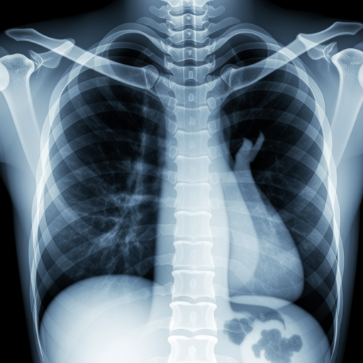
Figure 3: Feline thoracic radiograph demonstrating proper cardiac silhouette evaluation
Emerging Technologies and Future Directions
The future of veterinary radiography includes several promising developments. According to the Radiological Society of North America, artificial intelligence applications in veterinary image interpretation are advancing rapidly, with algorithms demonstrating 92-96% accuracy in fracture detection and 88-94% accuracy in pulmonary pattern recognition.
Other emerging technologies include:
- Cone-beam CT: Providing three-dimensional imaging for dental and orthopedic applications
- Portable Digital Systems: Enabling field radiography for equine and wildlife medicine
- Tele-radiology: Remote consultation services expanding access to specialist interpretation
- Dual-energy Radiography: Improved tissue characterization through material decomposition
Practical Considerations for Veterinary Practices
Implementing radiographic services requires careful planning. The AVMA Practice Facilities Guidelines recommend:
- Minimum room size of 3.5m x 3.5m for small animal radiography
- Lead-lined walls (1.5mm lead equivalence) for primary radiation barriers
- Dedicated electrical circuits with proper grounding
- Environmental controls maintaining 18-24°C temperature range
- Adequate storage for cassettes, grids, and positioning aids
Economic Considerations and Practice Integration
The financial aspect of veterinary radiography involves balancing equipment costs against clinical utility. A 2023 market analysis by Grand View Research indicates the global veterinary imaging market reached $2.1 billion, with digital radiography systems representing 38% of revenue share.
Return on investment typically occurs within 18-36 months for digital systems, considering factors such as:
Direct Revenue
Radiographic procedure fees and interpretation charges
Indirect Benefits
Enhanced diagnostic capabilities leading to increased treatment acceptance
Operational Efficiency
Reduced retake rates and faster patient throughput
Client Perception
Modern imaging technology enhancing practice reputation
Conclusion: The Evolving Landscape of Veterinary Imaging
Veterinary radiography continues to advance at an accelerated pace, driven by technological innovation and growing clinical demands. From basic fracture detection to complex three-dimensional reconstructions, X-ray imaging remains fundamental to veterinary diagnostics.
As digital technologies become more accessible and artificial intelligence integration expands, veterinary practitioners must maintain current knowledge through continuing education programs offered by organizations like the American College of Veterinary Radiology. The future promises even greater diagnostic precision, improved patient safety, and enhanced clinical outcomes through continued innovation in veterinary radiographic science.
References and Further Reading:
- American Veterinary Medical Association. (2023). Guidelines for Veterinary Radiographic Facilities.
- Thrall, D. E. (2018). Textbook of Veterinary Diagnostic Radiology. Elsevier.
- International Veterinary Radiology Association. (2022). Digital Imaging Standards in Veterinary Medicine.
- Bischoff, M. G., & Kneller, S. K. (2021). Radiation Safety in Veterinary Practice. Journal of Veterinary Medical Education.
