Canine Ventilator Management Techniques for Veterinary Professionals
Mastering Canine Ventilator Care: Essential Techniques for Pet Health Professionals
Ventilator support represents a critical intervention in veterinary critical care, particularly for canines experiencing respiratory failure. This comprehensive guide explores practical techniques, monitoring protocols, and troubleshooting strategies based on current veterinary respiratory medicine standards.
Understanding Canine Respiratory Physiology
Before operating any ventilator, veterinary professionals must appreciate the unique aspects of canine respiratory physiology. Dogs typically maintain a respiratory rate of 10-30 breaths per minute, with tidal volumes ranging from 10-15 mL/kg. The canine respiratory system differs significantly from human anatomy, particularly in bronchial branching patterns and lung lobe distribution.
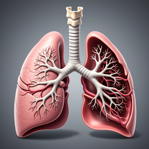
Anatomical illustration showing canine respiratory structures relevant to ventilator management
Ventilator Initialization Protocols
Proper ventilator setup begins with equipment verification. According to guidelines from the American College of Veterinary Internal Medicine, the following checklist should be completed before connecting any canine patient:
- Verify oxygen supply pressure (minimum 50 psi)
- Check ventilator circuit for leaks using manual breath delivery
- Calibrate pressure and volume sensors according to manufacturer specifications
- Set appropriate alarm parameters for the patient's size and condition
- Prepare emergency manual ventilation equipment
Mode Selection Strategies
Modern veterinary ventilators offer multiple operational modes. The Veterinary Information Network recommends the following mode selection criteria:
Volume Control Ventilation
Ideal for patients with stable lung compliance. Delivers consistent tidal volumes regardless of airway pressure changes. Recommended starting parameters: 10-15 mL/kg tidal volume, respiratory rate 10-20 breaths/minute.
Pressure Control Ventilation
Better for patients with variable lung compliance or risk of barotrauma. Maintains consistent peak inspiratory pressure. Initial settings: 10-20 cm H₂O PIP, I:E ratio 1:2 to 1:3.
Monitoring and Adjustment Techniques
Continuous monitoring represents the cornerstone of successful ventilator management. Capnography provides essential data about ventilation effectiveness, with normal end-tidal CO₂ ranging between 35-45 mmHg in most canine patients.
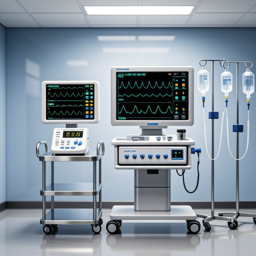
Modern veterinary monitoring station displaying ventilator parameters and physiological data
Key parameters requiring regular assessment include:
- Oxygenation: Maintain SpO₂ >95% and PaO₂ >80 mmHg
- Ventilation: Target PaCO₂ between 35-45 mmHg
- Hemodynamics: Monitor blood pressure every 15-30 minutes initially
- Patient-Ventilator Synchrony: Assess for signs of fighting the ventilator
Common Complications and Solutions
Even with proper technique, complications can arise. The American Veterinary Medical Association identifies several common issues:
Auto-PEEP Development
Inadequate exhalation time can lead to air trapping. Solution: Increase expiratory time by adjusting I:E ratio to 1:3 or greater, consider decreasing respiratory rate.
Ventilator-Associated Pneumonia Prevention
Elevate head 15-30 degrees, implement strict oral care protocols, and consider selective digestive decontamination in high-risk patients.
Weaning Protocols
Successful ventilator management includes appropriate weaning strategies. According to research published in the Journal of Veterinary Emergency and Critical Care, the following criteria should be met before weaning attempts:
- Resolution of underlying condition requiring ventilation
- PaO₂/FiO₂ ratio >200 on PEEP ≤5 cm H₂O
- Spontaneous breathing efforts present
- Hemodynamic stability without vasopressor support
- Normal neurological status (appropriate for patient condition)
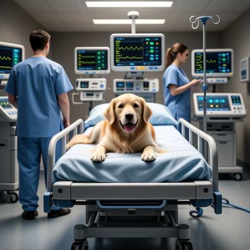
Veterinary team monitoring canine patient during ventilator weaning process
Equipment Maintenance and Safety
Regular maintenance ensures ventilator reliability. The FDA Center for Veterinary Medicine recommends:
- Daily circuit changes for patients on prolonged ventilation
- Weekly calibration of all sensors and alarms
- Monthly preventive maintenance according to manufacturer guidelines
- Annual comprehensive inspection and certification
Clinical Pearls from Experienced Practitioners
"Always have a manual resuscitation bag connected and ready during any ventilator adjustment. The 30 seconds it takes to locate emergency equipment can be critical." - Dr. Sarah Jenkins, DACVECC
"Document every parameter change, no matter how minor. This creates a valuable record for troubleshooting and improves patient safety." - Veterinary Technician Specialist, Critical Care
References: This article incorporates guidelines from the American College of Veterinary Internal Medicine, Veterinary Information Network, American Veterinary Medical Association, Journal of Veterinary Emergency and Critical Care, and FDA Center for Veterinary Medicine. Always consult manufacturer guidelines for specific equipment and institutional protocols for clinical practice.
